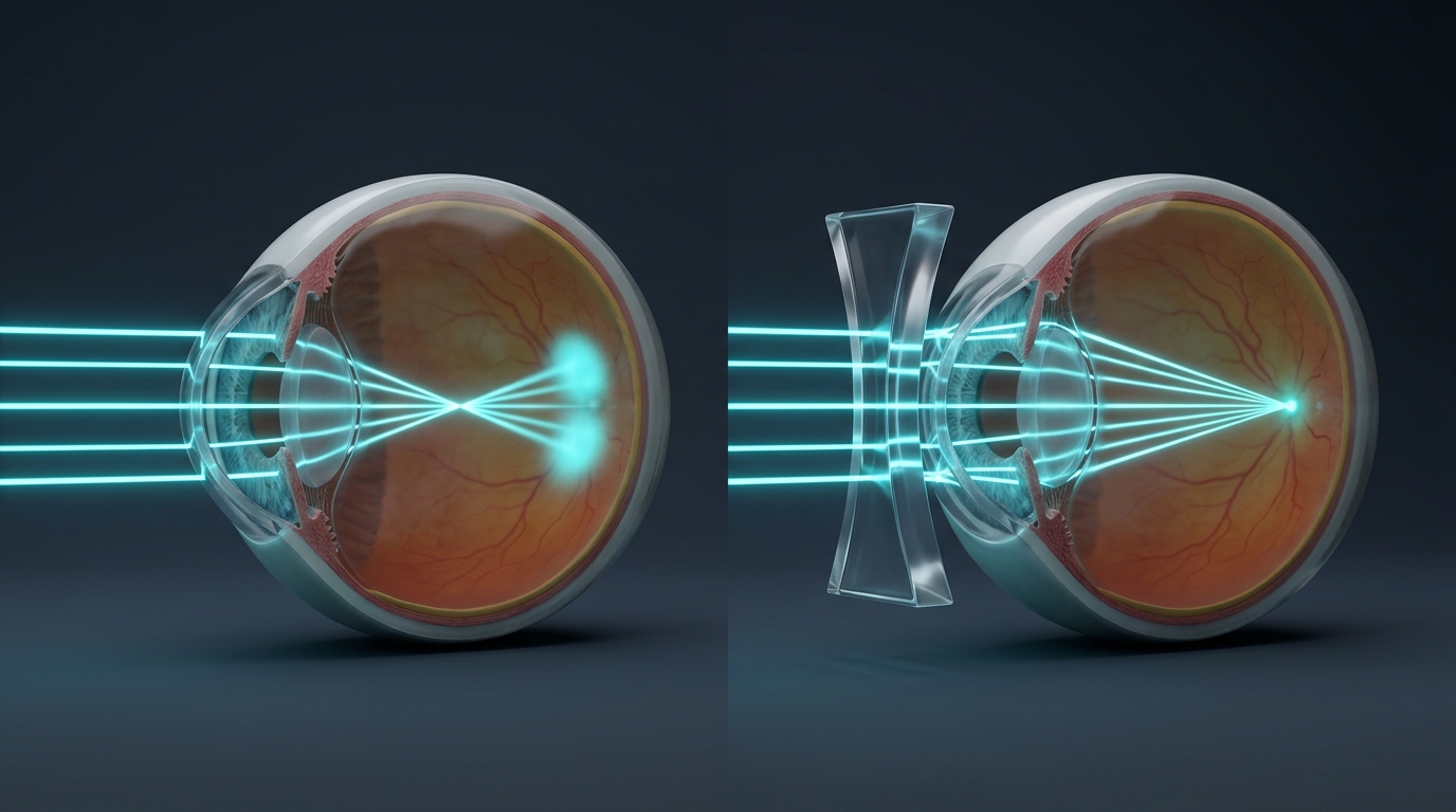
Anatomy of the Human Eye & Power of Accommodation
The human eye functions as a sophisticated organic camera that projects a real, inverted image onto a photoreceptor screen called the retina. The eyeball has an approximate diameter of $2.3\text{ cm}$.
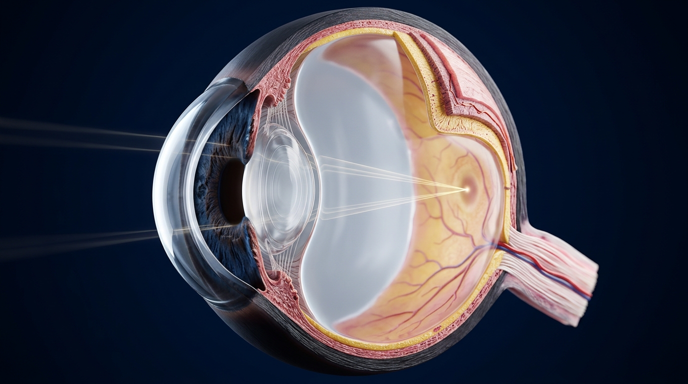
1. Key Eye Structures & Functions
- Cornea:
- Thin, transparent bulging membrane on the front surface of the eyeball.
- Crucial Optical Fact: Roughly $80\%$ of all refraction occurs at the outer corneal surface!
- Iris:
- Dark muscular diaphragm behind the cornea that regulates pupil diameter and gives the eye its color.
- Pupil:
- Central aperture regulating light admission (constricts in bright sunlight; dilates in dim lighting).
- Crystalline Lens:
- Flexible, fibrous double convex protein lens providing fine focal adjustments (accommodation).
- Ciliary Muscles:
- Smooth muscle ring adjusting lens curvature and focal length.
- Retina:
- Neural screen composed of millions of light-sensitive rods (dim light/intensity) and cones (color vision).
- Optic Nerve:
- Transmits electrical impulses directly to the visual cortex of the brain.
2. Power of Accommodation
- The physiological ability of the eye lens to adjust its focal length using ciliary muscles is called accommodation.
- Viewing Distant Objects ($u = \infty$):
- Ciliary muscles relax $\rightarrow$ Lens is pulled thin and flat $\rightarrow$ Focal length increases.
- Viewing Nearby Objects ($u = 25\text{ cm}$):
- Ciliary muscles contract $\rightarrow$ Lens bulges thick and curved $\rightarrow$ Focal length decreases.
3. Normal Vision Limits
- Near Point ($D$): The least distance of distinct vision without eye strain = $25\text{ cm}$.
- Far Point: The farthest point up to which the relaxed eye can focus = Infinity ($\infty$).
- Normal Range of Vision: $25\text{ cm}$ to $\infty$.